Having had a long-standing interest in insect macro photography I have often been tempted to take the next step and to examine and photograph microscopic life. However one of the main reasons I didn't was because of my erroneous belief that there was little scope for creativity in photo microscopy or much potential to take different styles of photograph.
Back in the Autumn I was very kindly given a book on microscopy which made me realise that, actually, there are a variety of ways to photograph microscopic life and plenty of scope for different lighting and other photographic techniques. Fast forward a few months and quite a lot of background reading and I became the proud owner of a 1960s Zeiss GFL microscope stand with a trinocular head (meaning a camera can be attached without having to remove the binocular eyepieces) and a selection of good quality Zeiss objective lenses. Although a vintage model (and older than I am!) the Zeiss GFL that I bought is in excellent condition, is superbly built and works as well as the day it was made. It also has very attractive 1960s styling which provides a nice contrast to my Olympus E-M1 which I can connect directly to it.
PLEASE CLICK TO ENLARGE IMAGES
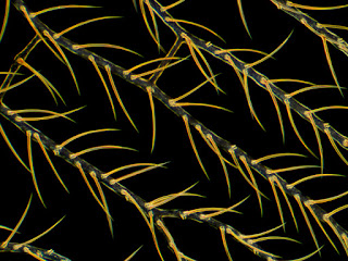
The hairy wing veins of a Green Lacewing (from an old prepared slide that I borrowed). Zeiss GFL, 100x magnification, dark field
A benefit of my Zeiss GFL is that it is capable of three different styles of image: (1) is 'standard' so-called brightfield where the image has a white background (2) is known as dark field where a black background is achieved and the subject is side lit which provides a very different image to bright field and (3) so-called phase contrast which provides a pale to mid grey background and the subject is normally shown with a lot of contrast which helps to pick out detail.
An iPhone image of my Zeiss GFL with trinocular head and Olympus OMD E-M1 attached. Note that I also connect the E-M1 to my iMac (which also sits on my desk) and can view the image directly on the iMac screen. This makes fine focusing much easier.
I have bought a number of prepared slides to view through the microscope, some, unbelievably, dating back to the 1860s, and have also made some of my own (temporary) slides viewing the life in standing water in my garden. It's a bit of a cliche but microscopy really does open up another world and I've very much enjoyed building my knowledge and skills over the last few months - though I still have a lot to learn.
Below are some of my favourite images taken so far.
Pine pollen composite, Zeiss GFL, 400x, darkfield, most were stacked
from 2-4 frames in Zerene Stacker (ZS)
A section of a young pine cone showing individual pollen grains, Zeiss GFL, 100x,
2 shots stacked in ZS, darkfield
An individual diatom (a type of microalgae with amazingly detailed shells). Zeiss GFL, 400x, Phase Contrast and stacked from 6 frames in ZS
An individual radiolarian (a type of microscopic protozoa, again with amazingly detailed shells). Zeiss GFL 400x Phase Contrast, 20 frames stacked in ZS
Filamentous algae Oedogenium. Zeiss GFL, 400x, Phase Contrast, 14 frames stacked in ZS
Pine pollen, Zeiss GFL, 400x, Phase Contrast, 4 frames stacked in Zerene Stacker (ZS)
Polypodium (fern) rhizome, Zeiss GFL, 160x, brightfield
A composite image of Diatoms. Zeiss GFL, 400x, darkfield, some diatoms stacked in ZS
A cross-section of a pine needle, Zeiss GFL, 100x, brightfield
finally, another single diatom, Zeiss GFL, 400x, Phase Contrast, 6 frames stacked in ZS


WOW!! Such an amazing work!! Congrats!
ReplyDelete